What is Ultrasound?
Human beings can normally hear soundwaves between 20Hz – 20000Hz. Ultrasound is sound waves with frequencies higher than the upper audible limit of human hearing. Ultrasound devices operate with frequencies higher than 20 kHz up to several gigahertz.
Physiologist Lazzaro Spallanzani was the first to study echolocation among bats in 1794, which forms the basis for ultrasound physics. In 1877 – Brothers Pierre and Jacques Currie discover piezoelectricity, which forms the basis of ultrasound transducers (probes). The first practical application of ultrasound is known to be during the World War I in detecting of submarines. Originally ultrasonic devices were used to detect objects & measure distances and later in several industrial applications such as mixing, cleaning etc.
Applications for ultrasound in medicine came about much later in 1930s and after and have become far more sophisticated.
Following categories of usage of Ultrasound devices in Hospitals are commonly seen:
1. Diagnostic
2. Therapeutic
3. Sterilization & Cleaning
Usage of Ultrasound for Diagnostic purposes
An ultrasound machine is used for abdominal, cardiac, maternity, gynecological, urological and cerebrovascular

examination, breast examination, small pieces of tissue as well as in pediatric investigations. An ultrasound device, consists of a transducer (or probe), transmitter pulse generator, compensating amplifiers, the control unit for focusing, digital processors and systems for display (Computer & monitor).
In the ultrasound diagnostics can be differed two techniques: transmission and reflection. Reflection technology (echo) largely in use today, registers the pulse as reflected from organs/tissues with different acoustic resistance. A piezoelectric transducer (mostly made of ceramic) encased in a probe usually sends and receives sound waves. The received data is processed and used to construct the image. The image is then a 2D representation of the slice into the body. 3D images can be generated by acquiring a series of adjacent 2D images. 2D phased array transducers are now used which can help process image faster and can even be used to make live 3D images of a beating heart.
Ultrasound frequencies in diagnostic radiology range from 2 MHz to approximately 15 MHz. Higher frequencies of ultrasound have shorter wavelengths and are absorbed/attenuated more easily. Therefore, higher frequencies are not as penetrating. This is why high frequencies are used for the superficial body structures and low frequencies are used for those that are deeper.
• 2.5 MHz: deep abdomen, obstetric and gynecological imaging
• 3.5 MHz: general abdomen, obstetric and gynecological imaging
• 5.0 MHz: vascular, breast, pelvic imaging
• 7.5 MHz: breast, thyroid
• 10.0 MHz: breast, thyroid, superficial veins, superficial masses, musculoskeletal imaging.
• 15.0 MHz: superficial structures, musculoskeletal imaging.
In diagnostic usage, ultrasound is usually produced in the frequency range of 2-15 Mhz, in very short, i.e., 1- to 5-cycle, pulses. Since the purpose of diagnostic ultrasound is detection of various organs and any abnormalities if any, the focus is on image capture. Therefore,temperature elevations and the potential for bio-effects are avoided, kept relatively low or negligible, through lower intensity and other factors described further.
Usage of Ultrasound for Therapeutic purposes
For therapy, ultrasound is used to induce heating of tissues and also through non-thermal mechanisms including ultrasonic cavitation, gas body activation, mechanical stress or others processes – to ablate tissues, fat or destroy tumour etc.
As against diagnostic purposes, in therapeutic applications of ultrasound lower frequencies are used generally between 20Khz – 5MHz. Therapeutic ultrasound devices may use short bursts or continuous waves to deliver effective ultrasonic energy to tissues. Ultrasound-induced heating is the result of the absorption of ultrasonic energy in biological tissue. As pressure amplitude, the frequency, or the propagation length is increased, the ultrasound wave can distort, which could ultimately lead to a discontinuity or shock in the waveform.
• Increasing frequency, nonlinear acoustic distortion, or pulse length can increase heating and enhance some non-thermal mechanisms.
• Decreasing frequency increases the likelihood of cavitation and gas body activation.
• Increasing power or intensity tends to increase the likelihood and magnitude of all bio-effects mechanisms.
Therapeutic Applications of Ultrasound Based on Heating
Physiotherapy – Unfocused beams of ultrasound for physical therapy were the first clinical application. This modality now typically has a base unit for generating an electrical signal and a hand-held transducer. The hand-held transducer is applied with coupling gel and moved in a circular motion over an injured or painful area of the anatomy to treat conditions such as bursitis of the shoulder or tendonitis, by trained physical therapy technicians. The object
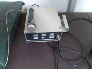
tive is to warm tendons, muscle and other tissue to improve blood flow and accelerate healing.
Hyperthermia – involves uniformly heating and destroy a tumor to about 42 °C for periods of about 1 hour, ultrasonically for the purpose of cancer therapy.
High intensity focused ultrasound (HIFU, or HIFUS) – In a HIFU system, a signal generator is connected to a focusing transducer, which produces very high local intensities of >1 kW/cm2 of 0.5–7 MHz ultrasound at the focal spot. The lesion produced in tissue typically may be a few mm in diameter and in length. The position of this spot must be carefully controlled and moved in order to ablate larger volumes of tissue. This method is used for treating uterine fibroids, visceral soft tissue ablation etc.
Therapeutic Applications of Ultrasound Based on Non-Thermal Mechanisms
Extracorporeal shockwave lithotripsy (ESWL) – is a widely used ultrasound therapy, which relies on nonthermal mechanisms. About 3000 shock waves are triggered at about 2 Hz repetition rate to pulverize kidney or gall bladder stone so that the pieces (<2mm) can pass naturally in urine. Shock wave devices similar to lithotripters are approved and marketed for orthopedic indications such as plantar fasciitis and epicondylitis
Phacoemulsification – A kHz-frequency ultrasound probe is used for phacoemulsification during surgery for cataracts. In phacoemulsification the eye’s internal lens is emulsified with an ultrasonic hand-piece and the lens debris removed by suction through the probe.
Low intensity pulsed ultrasound – has therapeutic application to accelerate the healing of bone fractures.
Ultrasonic cavitation – is used for liposuction etc.
Usage of Ultrasound for Sterilization and Cleaning
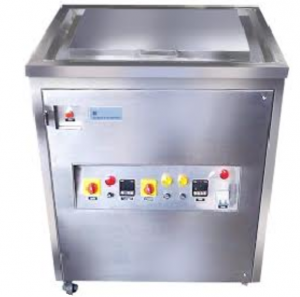
Ultrasonic cleaning is a process that uses ultrasound (usually from 20–400 kHz) to agitate a fluid. The ultrasound can be used with just water, but use of a solvent appropriate for the item to be cleaned and the type of soiling present enhances the effect. Ultrasonic cleaners are used to clean many different types of objects, including jewelry, lenses and other optical parts, watches, tools, coins, fountain pens, golf clubs, fishing reels, window blinds, firearms, car fuel injectors, musical instruments, gramophone records, industrial parts and electronic equipment.
In hospitals ultrasonic cleaners are used for dental and surgical instruments cleaning.