Laparoscopy is an operation performed in the abdominal or pelvic area using small incisions, typically less than 1.5 cm with the aid of a scope/camera, typically for surgeries on gall bladder, colon, kidney etc. Earlier, if any operation had to be performed in the abdomen, surgeons had to cut open the area – hence the operation was called laparotomy. A Laparotomy, naturally involves a big cut, more bleeding, pain, longer recovery time, higher chances of infection and on the whole more suffering for the patient. On the other hand, Laparoscopic surgery, also called minimally invasive surgery (MIS), is a relatively newer technique involving a smaller incision just enough to insert and use the scope and surgical instruments. Laparoscopic surgery is done using a laparoscope, essentially a rigid endoscope. What are the key components of a laparoscope? How does a laparoscope work? Read on to know more.
What are Laparoscopes and how are they used?
The laparoscope aids in viewing all the abdominal organs without having to cut open big and performing surgery on the relevant part without potentially damaging other parts of the body.
The smaller incision and minimally invasive operation helps in reduced bleeding and pain, results in shorter recovery time, reduced length of stay in hospital and reduced chances of infection.
Laparoscopes are nothing but rigid endoscopes, with a telescopic rod lens system, usually connected to a video camera – single-chip or three-chip. A digital laparoscope has a miniature digital video camera placed at the end of the laparoscope, replacing the rod lens system – but this is less popular.
To illuminate the operative area, so that the camera can capture clear images, a light source is used. A fibre-optic cable system is connected from the light source and inserted through the incision made via a 5 mm or 10 mm cannula / trocar. The abdomen is usually insufflated with carbon dioxide gas. This expands the viewing and operating space within the abdomen by stretching and lifting the abdominal wall away from the internal organs. CO2 is commonly used because the human body is very familiar with it and can be absorbed by tissues and removed through the respiratory system as usual. It is also non-flammable, an important feature since many electric devices are commonly used in operating rooms.
So what are the key components of a laparoscopy equipment system? How does a laparoscope work?
Key components of a Laparoscopy Equipment System
The basic laparoscopy equipment essential for any laparo-endoscopic procedure includes: endoscope, camera, light source, video monitor, insufflator, trocars and surgical instruments.
Trocar
A trocar is a device that is made up of a metal or plastic tip, a hollow tube called cannula and seal. During laparoscopic surgery, trocars are placed through the abdomen and just enough incision is made for this. The trocar then functions as a conduit for the subsequent introduction and retrieval of cameras and laparoscopic hand instruments, such as scissors, graspers, staplers, etc. Trocars are also used to aspirate gases or fluid from organs within the body, through a suction tube.
Light Cable
Flexible fibre-optic cables connect the light source, present outside, to the telescope/ laparoscope, so as to illuminate the areas to be examined and operated upon.
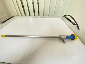
Laparoscope / Telescope
The components of a laparoscope are light post, light fibers, lens system, lens train, the shaft carrying the lens train, lens assembly at the proximal end and the eye-piece.
- Light post – The junction at which the fiberoptic light cable from the light source connects to the scope, to though light.
- Light fibers – Glass fibers carry light from the light post to the distal end of the scope, closer to the organs.
- Lens train – A series of glass rod lenses and spacers that transfer the image.
- Shaft – Stainless steel tube that houses the lens train and light fibers.
- Objective lens system – A collection of lenses, windows, and/or prisms located at the distal end of the scope, gathering the images. The distal objective can be manufactured at angles ranging from 0° to 120°, which enables the operator to see areas that might otherwise be out of view. Most common is the 00 and 300.
- Ocular lens assembly – The focusing lens of the scope located near the viewing end of the scope.
- Eyepiece – The eyepiece is located at the proximal. viewing end of the scope containing a magnifying glass. The image can be viewed through the scope, or the eyepiece can be attached to a camera coupler to view the image on an external monitor.
Telescopes can be 00, 300 or 450, with a diameter of 10mm and length 33cm. The 300 scope is preferred to 00 as it provides far greater latitude for viewing.
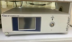
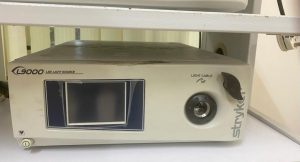
Light Source
The cold light source, halogen, xenon or LED may be used for laparoscopy. This maintains a constant colour temperature and allows automatic variation of the light intensity by the camera. The laparoscopy surgeons are trained in video laparoscopy which means performing surgery, while watching the image on a monitor screen instead of through-the-lens viewing.
Monitor
An HD medical grade monitor (15” – 32”) is part of the system for high quality images to be viewed during the procedure.
Laparoscopy Camera
Endoscopic camera systems that are high-definition (HD) or even with 4K image resolution are used to produce still and video images in the surgical field for laparoscopy procedures. The camera forms one of the most important components of the laparoscopy equipment. They are sensitive in the visible and infrared spectrum too. The optical image is transferred from the surgical site to the camera head by a variety of rigid and flexible scopes which are attached to the camera head. The system consists of a camera control unit (CCU) and a camera head with an in-built cable that connects to the CCU. The camera is usually equipped with a zoom lens allowing continuous adjustment of image magnification over a wide range. Lens aperture adjustment and white equalization would be automatic. Video output is available in different forms for viewing and printing etc.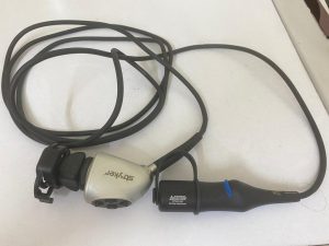
CO2 Insufflator
An Insufflator is used to deliver CO2 gas into the GI tract in laparoscopic examinations and operations. This expands the viewing and operating space within the abdomen by moving and lifting the abdominal wall away from the internal organs. CO2 is commonly used because the human body is very familiar with it and can be absorbed by tissues and removed through the respiratory system as usual. It is also non-flammable, an important feature since many electric devices are commonly used in operating rooms.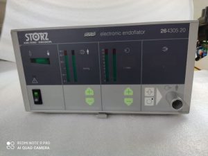
The insufflator sets and monitors required values for pressure, volume and flow of CO2 during the procedure. Patient safety circuits and alarms are usually built in.
Surgical Instruments
Surgical instruments used in laparoscopic surgery include forceps, scissors, probes, dissectors, hooks, and retractors etc.
Trolley
A trolley is typically used to house the entire ‘stack’ of laparoscopy equipment. The trolley or mobile video cart generally has a drawer and three shelves to hold all the equipment and related instruments. The upper shelves have a tilt adjustment and used for supporting the video monitor. The trolley is equipped with an electrical supply points on the rear side. The light source, video system & recorder and the insufflator can all be placed in mobile trolley. The drawer is used for keeping instruments, sutures, clips, staples etc. The trolley or cart is equipped with locking brakes and wheels.
Diathermy machine & lasers are also used in laparoscopy as necessary. Popular laparoscopy equipment brands are Stryker, Karl Storz, Smith & Nephews, Stork etc.
—————————————————————————————————————————
PrimedeQ is an e-Marketplace for buying, selling, renting, servicing and spares of medical equipment. We offer all types of used / refurbished medical equipment , including endoscopes, OT equipment, Laparoscopic Equipment, Lab equipment, X-Ray, TMT, ECG, anesthesia machine, ultrasound machines etc. We offer microdebrider, laparoscope , endoscope and ultrasound probe repair services etc.
https://in.linkedin.com/in/shanthi-mathur-ab07838