Both the Medical x-ray machines and security scanners are based on the same fundamental concept of using electro-magnetic radiation in the form of x-rays for scanning, imaging and detection. In case of security, it is the contents of the baggage and in case of medical X-Ray it is detecting abnormalities in the body part being scanned. The difference between security x-ray baggage scanners and medical x-ray scanners lies in the way image is captured and technology used for image manipulation required for relevant interpretation.
The visible spectrum of electromagnetic radiation has wavelengths from about 380 to about 750 nanometers. X-ray wavelengths range is much shorter, between 0.01 to 10 nanometers, not visible to human eyes. All x-ray images are produced in black and white based on how much X-Ray is absorbed by the object being scanned.
How is X-Ray used in security scanners and medical scanners?
All medical X-Rays are in various shades of gray, black and white. This is because different tissues in the body absorb different amounts of radiation. Bones in our body absorb most of the x-rays , hence the least amount of radiation through bones reaches the detector. Therefore the bones look white in the X-Ray image. Muscles, fat and other soft tissues absorb less and so appear gray. Cavities look darker. X-Ray will completely pass through air. This is the reason any fractures in the bones shows clearly black.
Medical X-Ray images can be taken from different angles and perspectives to form an opinion. In some cases such as Flouroscopy and in some CTs contrasts and dyes can be used to produce studies with contrast. In case of CTs, multiple slices are produced per second resulting in 100s and 1000s of images to construct a 3D image. Radiologists are trained to process and interpret all these types of images in medical x-rays. Most often the images need to be clinically correlated to arrive at a diagnosis and further course of treatment.
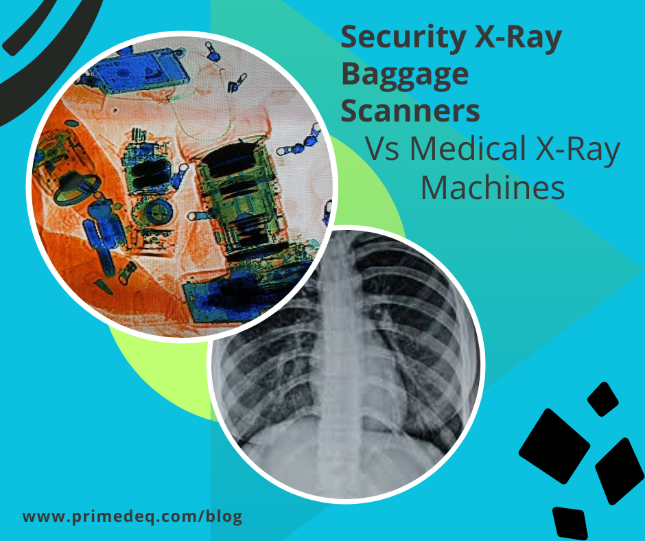
In case of Security X-Ray baggage scanners, just black & white images are not adequate because the scanned objects are not only organic, but could be inorganic or mixed. Therefore, software applications are used for colouration of the images, based on their atomic numbers. The material is identified using their atomic numbers. As per Moseley’s Law the square root of the frequency (ν) of characteristic X-rays emitted by an element is directly proportional to its atomic number (Z), represented by the equation ν = a(Z – b), where a and b are constants. Generally organic materials (plastics, food, etc.) are coloured orange, inorganic materials like heavy metals (steel, gold, lead, etc.) are coloured blue and items with a combination of organic and inorganic material such are shown as green in the image. Brightness of the colour depends on the thickness of the material. A thicker material will appear darker.
Security personnel use both the black and white images and the coloured images depending on the case to identify objects. Some security x-ray baggage scanners may provide dual images, vertical and horizontal and some are capable of providing 3D view of the scanned baggage.
Using all these images from different angles and colours, security personnel must very quickly identify what a baggage contains and spot suspicious objects that may even be hidden inside another – all within a few minutes. Speed is of essence due to large volumes of baggage and passengers in setting like airport etc.
What are the key technical differences between baggage scanners and medical x-ray machines?
- mA ratings & Levels of ionizing radiation – Security X-Ray baggage scanners typically use low milliamperes (mA) and a fixed voltage for their X-ray operation, meaning the voltage is not varied during the scan. The current is usually in the range of 0.1 mA to 1.2 mA, with voltages between 100-160 kilovolts (kV). These scanners utilize low levels of ionizing radiation, which is adequate to create the images needed to check the luggage contents. Medical X-Ray machines on the other hand use 20mA – 800mA. In addition, depending on the study required and the patient age & size, the voltage and exposure time would are varied. The ionizing radiation involved in baggage scanning is as low as 1mSv, almost same as the atmospheric levels. However in medical x-rays it could be as high as 25mSv in CT/ PET Scans etc.
- Image processing – As described in the previous section, the ways in which the X-Ray images obtained is manipulated using atomic number of the elements, colouring the images based on organic or inorganic and obtaining dual or 3D imaging. Medical imaging manipulation includes using contrast/dyes and multiple slices as in CT scan to build a 3D image, but all of it in grayscale.
- Radiation safety protocols – All types of X-Ray and Gamma radiation equipment need approvals from AERB. Radiation levels being high in medical x-ray systems, the regulatory and safety protocols are very stringent. Security x-ray baggage scanners use what is called a Cabinet X-Ray Systems. This type of system contains an x-ray tube installed within a shielded enclosure. The enclosure is made up of a material, usually lead, that prevents most of the x-ray radiation from escaping the cabinet. The baggage items are passed through this enclosure via a conveyor belt. In case of medical x-ray machines, the entire room/ door etc. are made with lead lining. In addition the technicians use lead aprons and stand behind a protective lead shield, while exposing the patient.
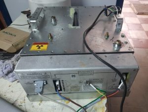
Repair of Security X-ray scanners
A baggage x-ray scanner consists of several components such as:
- X-ray Source
- Detector
- Image Processing Unit
- Control Console
- Display Screen
- Radiation Shielding
- Conveyor Belt
- Alarm System etc.
While all other components may be built differently, the core X-Ray source technology is the same. Therefore repair of X-Ray tube is done the same way. If repair of x-ray tube in a security x-ray baggage scanner is required, PrimedeQ can assist you.
For key components of a medical x-ray and its working see https://www.primedeq.com/blog/how-does-an-x-ray-machine-work-what-are-the-main-components-of-an-x-ray-unit/
The technology behind the X-Ray source i.e. the X-Ray Tube is the same for medical applications. For more information on X-Ray machine repairs check out the following blogs:
—————————————————————————————————————————
PrimedeQ is an e-Marketplace for buying, selling, renting, servicing and spares of medical equipment. We offer all types of used / refurbished medical equipment , including used X-Ray or C-Arm machines, ultrasound machines and surgical equipment, endoscopes, OT equipment, Laparoscopic Equipment, Lab equipment, TMT, ECG, anesthesia machines etc. We offer repair services for all types of medical equipment including Lab equipment, Ophthal equipment such as AR, slitlamp, ENT equipment like microdebrider, laparoscope , x-ray tube, rigid and flexible endoscopes as well as EUS (Endoscopic Ultrasound Units) and ultrasound probe etc.
Contact us on +91 8971223957/ 7019759765 or [email protected]
https://in.linkedin.com/in/shanthi-mathur-ab07838