Many of our YouTube subscribers ask for the mA KVp chart. Often young X-Ray technicians are put to work without any orientation to the new X-Ray machine and certainly with no exposure chart to serve even as a starting point. Not knowing what exposure settings to use when a patient has presented himself/ herself and repeating X-rays to get the right image, can be very frustrating when time is limited, especially with unwell and not-so-cooperative patients. By having a good exposure chart that is easy to refer to and simple to use – one can greatly reduce the number of repeated exposures and make taking x-rays simpler.
A good quality exposure chart is one of the most important tools to help a radiographer achieve high quality X-ray images, reduce the number of repeated X-rays and as a result the radiation dosage to the patient.
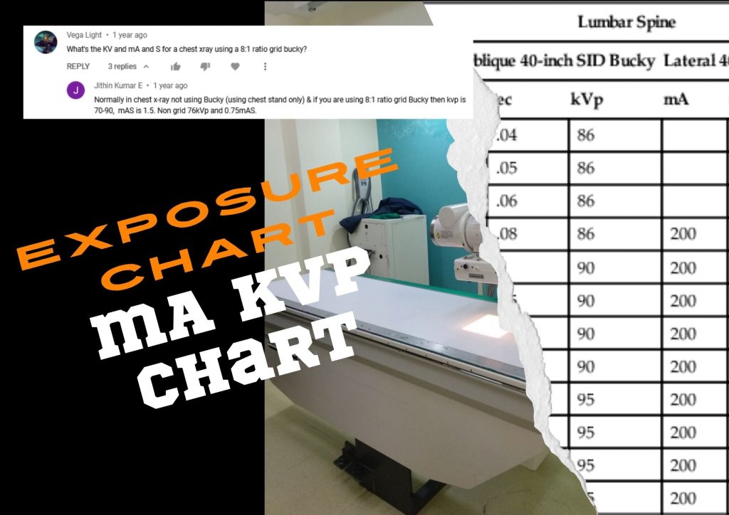
What are Exposure Chart or mA KVp chart?
An exposure chart or technique chart is a listing of the various radiographic examinations along with respective exposure factors. Most mA KVp charts are based on selecting the body part to be scanned and the thickness of that body part. Against the type of examination and patient/part measurement (in centimeters), an exposure chart ideally includes kVp, mA, exposure time, projection, Source-to-Image Distance (SID) and a grid (Bucky) notation.
The reason for starting with body part and its thickness is that you need different kV and mAs settings for say, a 10 cm thick thorax than you do for a 10cm thick skull. Denser organs like bones need higher kV to penetrate it. Similarly, a thorax and an abdomen would need different settings as the Thorax contains a lot more air and less soft tissue than an abdomen. Also a Pelvis ideally needs more kV to adequately penetrate through it, in comparison to the abdomen.
Historically, especially for conventional line-frequency x-ray machines, many technicians relied on standard mA and KVp figures for each part being exposed for an average sized patient. If the patient seems significantly different sized from average, then tweak the mA and KVp settings upwards or downwards, suitably, and hope the exposure was right.
However, exposure charts are not the same for all x-ray machines. They are specific to each x-ray machine and each facility. The x-ray machine manufacturer cannot supply a definitive chart with the machine because the exposures will vary depending on the types of grids, table tops, and SID at the hospital facility. This is why you cannot pick up exposure chart of any machine and start tweaking it. It is important to know what to change when setting up your exposure chart, and what to look at on the resulting images to enable one to tweak the chart to best suit each facility’s needs.
A slight over exposure will give a better quality image than an under exposed image, but it is always best to aim to get as close to fine tuning as possible so that we do not end up over-dosing patients.
So, what are the various factors to consider?
What are the various exposure factors?
The primary exposure technique factors the radiographer selects on the control panel are milliamperage (mA), time of exposure, and kilovoltage peak (kVp).
However there are other factors to consider, so as to obtain proper image.
Source: Exposure Technique Factors | Radiology Key
- Primary Factors
- Milliamperage and Exposure Time
- Kilovoltage Peak
- Secondary Factors
- Focal Spot Size
- Source-to-Image Receptor Distance
- Object-to-Image Receptor Distance
- Calculating Magnification
- Central Ray Alignment
- Grids
- Beam Restriction
- Generator Output
- Tube Filtration
- Compensating Filters
- Patient Factors
- Body Habitus
- Part Thickness
- Pediatric Patients
- Special Considerations
- Projections and Positions
- Casts and Splints
- Pathologic Conditions
- Soft Tissue
- Contrast Media
Creating an exposure chart for your facility
When a new chart is being created, the best way to do it is:
- Ask the x-ray vendors to supply a standard chart relevant to their x-ray machine.
- He/she can be asked to fine-tune it for your facility too, depending on the grid and SID etc., at the time of installation.
- An experienced radiology technician from the hospital team could update this mA KVp chart further for the type of patients and procedures being done in the facility, for all technicians to use.
This is the best way because the chart would be tailor-made specifically for the facility as well as the equipment, using only settings available on your control panel.
Important note: An exposure technique chart should be prepared with a calibrated x-ray machine and the specific type of image receptor (IR) used. If there are any changes, it is time to revisit the mA KVp chart.
Exposure technique for HF or automatic exposure control X-Ray machine
The conventional X-ray system cost varies from Rs. 1 lakh to Rs.5 lakh, but is old technology with limitations on image quality & radiation dose control. The fully digital X-ray systems offer excellent imaging solutions but are expensive (Rs 25 lakhs to 1.5 crore plus) and unaffordable for most hospitals in India.
Line-frequency X-Ray machines are being replaced by High Frequency x-ray machines. Most HF X-ray machines use automatic exposure control (AEC) programmed for each anatomy being scanned. The HF x-ray machines are better technology and more automated offering several advantages over line frequency machines.
Advantages of HF X-Ray machines
- Consistent image quality – The radiographic quality is consistently good. There is good uniformity across the image.
- Lower exposure time/ radiation dosage – The exposure factors required, are 20%+ lower than that required by the 2-pulse conventional generator.
- The fast and quick user interface – The console’s digital display and anatomical programs make imaging extremely easy while reducing time and exposure. This also helps in increasing patient throughput.
- Higher x-ray generator power – With the HF technology, the radiographic output is in the range of 30 to 80 KW. Higher power generator increases ability to do quicker scans even for paediatric patients and trauma patients, where a short exposure time is preferable.
- Floating radiographic table – The floating table top provides easier up & down movements and tilting, making on-boarding as well as positioning of patients of all types much easier.
- The HF X-Ray systems creates a safer imaging environment by reducing exposure time, lowering radiation dose by as much as 25% and eliminating soft radiation compared to conventional X-rays.
Setting the exposure factors in HF X-ray machines
The Control Console is an important component of an X-ray generating system. There are three main adjustable controls that regulate the 1) tube voltage in kilovolts, 2) the tube amperage in milliamps, and the 3) exposure time in minutes and seconds. Some systems also have a switch to change the focal spot size of the tube.
HF X-Ray machines today have digital console with settings for procedure being done (e.g AP Lumber) and the patient thickness, rather than the Voltage, Current and Exposure time. These exposure factors are automatic (pre-programmed) based on the procedure and patient thickness setting, making it easier for the technician.
Does this mean exposure charts are not needed in case of HF x-ray machines with automatic exposure control?
The answer is – no. Automatic exposure control or anatomically programmed radiography systems must also have a technique chart. The chart should indicate all the items that a manual technique chart does, except the exposure time (which is automatic).
HF X-Ray Systems are useful as they take care of the Primary exposure factors and some of the secondary exposure factors for an average patient. But they are still not good for all types of patients. Exposure techniques vary from equipment to equipment. Hence the hospital still needs an exposure chart for all technicians to follow. However HF machines could offer a good starting point to start building an exposure chart from.
——————————————————————————————————————————
PrimedeQ is an e-Marketplace for buying, selling, renting, servicing and spares of medical equipment. We offer all types of used / refurbished medical equipment , including maternity equipment, Lab equipment, X-Ray, TMT, ECG, anesthesia machine, ultrasound machines, endoscope and ultrasound probe repair services etc.
https://in.linkedin.com/in/shanthi-mathur-ab07838