A-scan machine is essentially a diagnostic ultrasound device that is used for ocular biometry, i.e. measuring dimensions of various parts of the eye. The various parts of the eye measured by A-scan include corneal curvature, axial length, and anterior chamber depth and can be useful in diagnosing common eye problems. A-scan machine is primarily used by ophthalmologists to determine the power of the intraocular lens (IOL) needed for implant in cataract surgery. This blog is about how A-scan works, what are the major components and most common A-Scan repair issues observed.
A-Scan biometry techniques
Ocular biometry using A-scan may use one of two techniques – Direct contact or Immersion.
- Direct contact technique requires that the ultrasound probe be placed directly on the corneal surface, flattening it, using an ultrasound probe. This method is hence called applanation ultrasound biometry.
- In Immersion biometry, a small fluid shell (immersion shell) is placed on the anesthetized corneal limbus. The limbus forms the border between the transparent cornea and opaque sclera, contains the pathways of aqueous humour This is the site of surgical incisions for cataract and glaucoma. The A-scan probe is held within the fluid, thereby avoiding direct contact with the cornea surface.
Many A-scan machines offer both techniques. Popular A-Scan machine models are, A-Scan G-Matronix, Matrix, Biomedix Echorule, Sonomed Pacscan, Keeler Accutome, Souer A-scan, Quantel Medical Axis Li etc.
How does an A-Scan machine work?
A-Scan ultrasound probe/ transducer
A-scan biometry, uses a thin ultrasound probe to scan the eye. The probe is used to generate a sonic beam generally at a frequency of approximately 10-11 MHz and receive the echo back from each interface in the eye. An interface is the junction between any 2 media of different densities and velocities, such as the anterior corneal surface, the aqueous/anterior lens surface, the posterior lens capsule/anterior vitreous, the posterior vitreous/retinal surface, and the choroid/anterior scleral surface.
Echoes received back into the probe from each of these interfaces are converted by the biometer to signal spikes arising from baseline. The greater the difference in the 2 media at each interface, the stronger the echo and the higher the spike. Obviously the ultrasound probe is the most important component of the A-scan machine and the most frequent A-scan repair issue is problems with the probe.
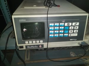
Control Unit & accessories
A control unit helps perform the operation and receives the results. The data as received is displayed either on the display screen of the A-scan unit or laptop/computer depending on the model. The various controls typically include the support for contact or immersion, independent velocities, automatic capture, automatic alignment indicators, gain control, and manual and automatic capture settings etc. A foot switch is used to stop, start and review the axial length measurements. A printer either connected to the computer or display unit is required to print results.
All ultrasound devices have gain control. The gain control is a knob, button and/or a series of sliders on the console, and is used to adjust scanning parameters. Gain control is not just an image brightness adjuster. Gain ensures appropriate ultrasound signal amplitude when the signal is returning from the tissue to the transducer at its output, despite variation of the signal amplitude at the input. In A-Scan machine typically a potentiometer helps with these setting adjustments. Issues with potentiometer are another common A-scan repair problem users face. Fixing this can be an urgent need.
Key Components of A-Scan machine
The key components of a typical A-Scan machine are: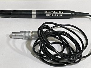
- Ultrasonic probe
- Control Unit
- Footswitch
- Laptop or computer or display (depending on the model)
- A-Scan Software
- A-B Type USB Cable to connect from control unit to computer
- Printer to print results
- Immersion Shell – Optional
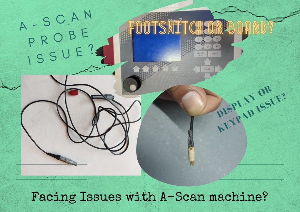
Major A-scan Repair or Service issues
The majority of service issues in A-Scan machine concern the major components mentioned above. The most common A-Scan repair issues we handle are: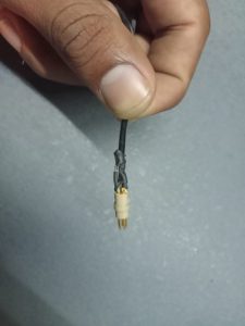
- A-Scan Probe repair & replacement
- Potentiometer repair or replacement
- Display issue
- Printer issue
- Power supply board repair or replacement
- Mother board repair or replacement
- Key pad issue
We offer repair services for all types of medical equipment including A-Scan repair service, microdebrider, laparoscope , rigid and flexible endoscopes as well as EUS (Endoscopic Ultrasound Units) and ultrasound probe etc.
PrimedeQ is an e-Marketplace for buying, selling, renting, servicing and spares of medical equipment. We offer all types of used / refurbished medical equipment , including A-Scan machine and other Ophthalmology equipment, endoscopes, OT equipment, Laparoscopic Equipment, Lab equipment, X-Ray, TMT, ECG, anesthesia machine, ultrasound machines etc.
https://in.linkedin.com/in/shanthi-mathur-ab07838