X-Rays and Ultrasound scans are two of the most common medical imaging investigations used to see inside the body and diagnose diseases, injuries etc. What is the difference between X-Ray and Ultrasound scans? How are the two medical equipment different in terms of technology they use? Are they used for the same purposes or different investigations? How do X-Ray and Ultrasound machines compare in terms of cost, patient safety, ease of use and other parameters? In this blog we talk about key differences between X-Rays and Ultrasound machines in terms of technology used, appropriate applications, safety, cost and which investigation is likely be prescribed by the doctor based on the organ or problem area to be examined and other relevant factors.
Difference between X-Rays and Ultrasound in technology
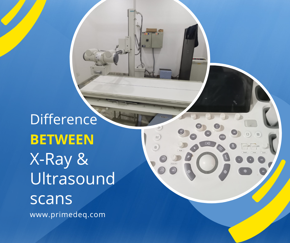
Ultrasound scanners use sound waves, while X-ray machines use X-radiation to scan and generate images.
Ultrasound technology
A diagnostic ultrasound machine uses high-frequency sound waves to perform scans.
Ultrasound essentially means sound waves with frequencies higher than the upper audible limit of human being, e,g, 20KHz.
Diagnostic ultrasound machines operate with frequencies ranging from 2 MHz to approximately 15 MHz. High frequencies are used for the superficial body structures and low frequencies are used for to examine deep-seated organs.
Ultrasound scans include a hand-held transducer pressed close to the skin of the body part to be scanned. This transducer transmits as well as receives the high-frequency sound waves. A sonologist moves the transducer around to generate sound waves from different angles. Sound waves reflect off the various tissues. The transducer is interfaced with a machine which records and processes the echoing waves, and translates them into images on a monitor screen. A console is provided with knobs to vary the settings for suitable scans. The images processed may be stored in the computer or printed out from attached printer.
How are images generated?
The sound waves are reflected back to the transducer from the interfaces of tissues, fat, fluids, bones etc. at the border/edges of the organs in the path of the wave (e.g. the changeover between fluid and soft tissue or tissue and bone) due to their varying densities. Sound waves penetrate fluids and softer tissues more easily and reflect less, while bones and air do not transmit sound and hence reflect fully. Based on the speed and amplitude of sound and the time of each echo’s return, the scanner estimates the distance from the transducer to the tissue. These calculations help generate two-dimensional images of tissues and organs.
As mentioned earlier sound does not travel much through air or dense structures like bones and teeth. Hence ultrasound imaging is not used to detect fractures etc. However, since it penetrates through fluids, fat and soft tissues well, it is used to scan organs not shielded or blocked by bones such as abdominal organs. Ultrasound can detect and define the size, shape of soft tissue organs, identify fluid presence in places it should not be and identify tumours etc., by abnormal thickness of tissues.
X-Ray machines technology
An X-Ray machine consists of two main components – X-Ray generator and an image detection system. X-radiation is produced in an X-Ray tube, consisting of cathode and anode. These X-Rays are focused by a collimator on to a photographic plate, where the image is captured. The patient is placed in between the X-Ray tube and the photographic plate. E.g for chest X-Ray the plate is on a stand behind the patient. For hip x-ray the patient lies on a bucky table under which the photographic plate is placed to capture the image and the x-rays come from the tube above the patient. The X-rays penetrate through different body parts in varying degrees based on their density and thickness, and reach the photographic plate. The x-ray image thus created is black & white with various shades of grey based on varying tissue density. X-Rays pass easily through air, soft tissue, like organs and muscles, but not so easily through hard tissue like bones and teeth. Low density material such as air does not absorb x-rays hence appears black. Dense materials such as metals /metal based contrast medium absorb x-rays heavily and hence appear bright white. Bones and teeth also appear almost white due to their high calcium content. All other soft tissues, fluids, fat etc, appear in various shades of grey.
Abnormality is detected if there is any unexpected increase or decrease in the density of a known anatomical structure noticed.

It is harder to distinguish between fluids, fat and soft tissues in an x-ray, because the contrast in the greyness is not much. However the contrast between air and bones and teeth is stark, due to which a fracture in a bone filled with air would show clearly. Hence X-rays are most commonly used to scan skeletal structures. This is because bones e.g. have calcium, a metal. For several investigations such as abnormality of GI tract, patient may be asked to ingest a Barium compound that will fill a space of interest and show up as white in X-ray imaging.
Difference between X-Rays and Ultrasound in Application
Ultrasound Applications
The most popular use of ultrasound machines is during pregnancy to monitor fetal development or detect genetic disorder if any.
https://www.primedeq.com/blog/top-4-ultrasound-tests-to-know-during-pregnancy/
Ultrasound can be used to create images of muscles, joints, and tendons, reproductive organs, bladder, thyroid, gallbladder, spleen, and pancreas. An ultrasound scan helps in diagnosing cardiovascular issues, abdominal issues, infections, tumors and cysts, and soft tissue injuries.
One of the most prolific applications of ultrasound imaging is in echocardiography. Echocardiography is examination of the blood flow, cardiac chambers, and functions of the heart and valves. Doppler effect in ultrasound waves plays a significant role in identifying heart diseases.
Ultrasounds provide far more details about the soft tissues even in the musculoskeletal system than an X-ray report. Ultrasound can easily identify any tears in the ligaments and tendons of muscles due to unexpected changes it detects in the density of these cells. Ultrasound can tell us the degenerative as well as healing state of soft tissues which X-Rays cannot. In other words Ultrasound provides more details than X-ray scans.
However in certain areas Ultrasound has its limitations. As sound waves are fully reflected off air or bone, it isn’t effective at imaging body parts that have gas in them or are hidden by bone, such as the lungs or head. Ultrasound may also be unable to discern deep-seated parts of the body, as it may not be able to penetrate that far. To view these areas, other diagnostic imaging techniques such as CT or MRI may be recommended.
X-Rays Applications
X-rays are most effective at creating images of hard tissues, such as bone and teeth. The bone shows up as white in an X-ray image due to its calcium content with the overall image in black and white. Air, which does not absorb x-rays well, shows up as black in an X-ray image. The contrast in colour between bones and air helps with investigations of organs like lungs. Chest X-rays are perhaps the most common x-ray examination. It can reveal enlargement of the heart, fluid buildup, and lung infections, tuberculosis, and some forms of cancer. X-rays are also most commonly used in diagnosing fractures. X-ray technology is used in DEXA bone density scans and mammograms hence helping with diagnosis of osteoporosis and breast cancer. Dental X-Rays are used to widely to examine teeth and gums.
Barium along with standard X-ray imaging is used for investigations of Gastro-Intestinal (GI) tract. Barium sulfate is a radiopaque contrast media, which absorbs x-rays and appears white on X-ray film. When ingested into the GI tract, barium coats the inside wall of the esophagus, stomach, large intestine, and/or small intestine so that the inside wall lining, size, and shape are all clear on X-ray. This ensures abnormalities of the GI tract, such as tumors, ulcers and other inflammatory conditions, polyps, hernias, and strictures can be clearly seen by x-ray examination. Similarly, air-contrast or double-contrast GI studies are also done.
Barium studies are usually done in Fluoroscopy mode rather than radiography mode of X-Ray. In Fluoroscopy mode a continuous stream of X-ray is passed through the body part being examined resulting in a stream of x-ray images captured like a video. This allows the radiologist to see the flow of the barium through the GI tract. When there is an abnormal growth, which constricts the flow, it immediately becomes visible.
Patient safety, Cost and other considerations
Excess exposure to X-ray radiation could be harmful both to patient and caregivers. Hence X-Ray usage is highly regulated while ultrasound is considered safe. This is a significant difference between X-Rays and ultrasound scans. X-Rays are not considered safe for pregnant woman, while ultrasound is most prolific in use during pregnancy.
Cost wise, basic conventional high-frequency X-Ray machines and basic ultrasound machine cost about the same. However, high-end ultrasound machines are extremely feature rich and may cost 3-5 times more.
In India X-Ray machines use is regulated by AERB. A valid operating license is required to operate x-ray machines. Ultrasound operation is not harmful in itself; however a PCPNDT license is required in India.
—————————————————————————————————————————
PrimedeQ is an e-Marketplace for buying, selling, renting, servicing and spares of medical equipment. We offer all types of used / refurbished medical equipment , including ortho drills and other surgical equipment, endoscopes, OT equipment, Laparoscopic Equipment, Lab equipment, X-Ray, TMT, ECG, anesthesia machine, ultrasound machines etc. We offer repair services for all types of medical equipment including Lab equipment, microdebrider, laparoscope , x-ray tube, rigid and flexible endoscopes as well as EUS (Endoscopic Ultrasound Units) and ultrasound probe etc.
Contact us on +91 8971223957/ 7019759765 or [email protected]
https://in.linkedin.com/in/shanthi-mathur-ab07838