
How to decide which C-Arm machine to buy? The key features to consider when buying a C-Arm machine include – x-ray generator, tube type & collimator system, C-arm dimension, maneuverability, and range of movements, FOV, compactness required, image processing, storage and transfer capability, radiation dosage, image detection and capture technology etc.
Choice of type of C-Arm depends on what procedures it is going to be used for. C-Arms are used in a range of different gastrointestinal, peripheral vascular, neurosurgery, pain management & Anesthesiology, urology, orthopedics and a few other procedures in like HSG, varicocele embolization in Operation Theaters today. Specifications would differ for different types of procedures. For e.g for short needle placement procedures for pain management a stationary anode may suffice. For longer procedures that are frequently done, rotating anode may be required. Again, whether II with FOV or 9” is appropriate or is 12” required depends on the type of procedures. While 12” C-arms may be larger and costlier, they are more suitable for vascular studies.
Every hospital works with budgetary constraints. Hence complete clarity on its proposed use (i.e. what types of procedures) is critical in selection of C-Arm machine.
When buying mobile C-arms, some of the basic considerations are as follows:
X-ray generator
With regard to X-Ray generator in a C-arm machine, there are three main points to be looked at:
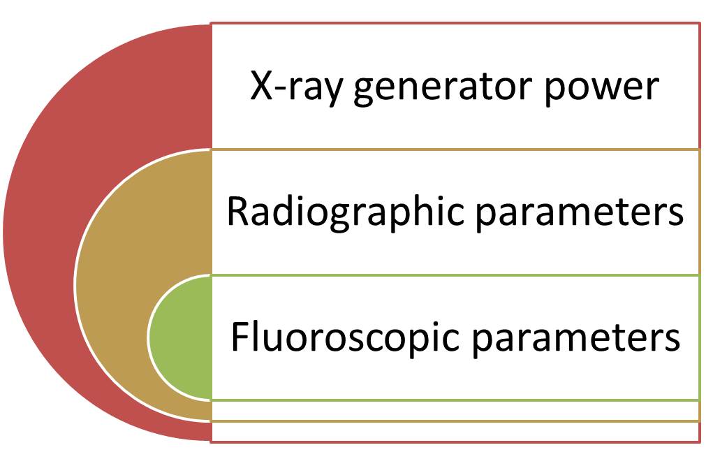
X-Ray generator Power
The benefit of increased x-ray power is that it allows greater quality of imaging with shorter exposure times. It also reduces risk for error and re-work.
C-Arms come with a range of power options anywhere between 2.5 KW – 25 KW. Some of the basic c-arm machines like Siemens Multimobil 5C (2.5KW), Allengers HF-49 (3.5KW), Skanray SkanC (3.5 KW) or Philips Surgico 60D HF (2.5KW) are in the lower power range. While models like Allengers HF 59R (15KW), latest Siemens machines like Cios Alpha and Cios Spin etc. (25KW/ 15KW), OEC Elite CFD (15KW) offer far more power.
Flouroscopy & Radiography Modes
C-Arms have both radiographic and fluoroscopic options. Most of the basic C-arms come with atleast two fluoroscopic modes – ADR (Automatic Dose Regulation) and manual modes. Recent models feature many other modes apart from the two above, like
- Pulsed fluoroscopy,
- Continuous high fluoroscopy,
- Digital Subtraction Angiography,
- Digital cine mode etc.
Dimensions, Maneuverability and Range of movements
C-arm units are increasingly sought to be light-weight, easy-to-move, compact and efficient. Hence the dimensions and weight of the unit are important. Very old C-Arm machines used noisy AC motors for facilitating movements, while newer ones use noiseless DC motors.
The following parameters are important among features to consider when buying a C-Arm machine – these are normally compared between C-Arms in terms of movement capability:
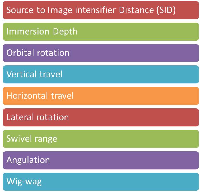
C-Arm Depth, Rotation and Movement capabilities
A great C-arm depth may be preferable, but it should not be too large making it difficult to move around in the OT. Source to II distance (SID) in C-Arms now ranges anywhere between 86cm to as high as 100 cm. Most basic C-arms would have an orbital rotation capability of 1250 (overscan/underscan – (-350/+900), some older ones may have lesser ~1150. However some recent C-Arms like OEC Elite (1450) and Siemens Cios Alpha (1480) orbital rotation and Siemens Cios Spin boasts of 2000 (-/+ 100) rotation.
The C-arm must be deep enough to accommodate all sizes of patients and low enough to fit underneath the hospital’s beds and operating theatre tables. At the same time it should be easy to rotate as much as required to align with the patient without hitting something. Hence from the surgeon’s point of view – Orbital, horizontal, vertical and lateral movements, angulation and swivel range are all important considerations.
Latest in C-Arm movement
In terms of C-Arm movement there are two cutting edge features being talked about:
- SmartView pivot-joint as seen in GE OEC Elite 9900 SmartView. Having the best image quality is of no use if surgeons do not get the perfect alignment. Perfect alignment often requires great effort on the part of the care-givers, too many movements of the machine and patient re-positioning, in turn increasing exposure time and radiation dose, very often not even achieving desired results. A pivot joint makes the movements and alignments smooth, easy and finely calibrated.
- Isocentricity – As seen in Siemens Cios Spin – isocentric C-arms move in a perfect circle around the subject while other models rotate in an oval pattern. For a C-arm to be “isocentric”, the central x-ray beam must remain in the isocenter of the subject regardless of the position of the C-arm. The distance of the X-ray tube and the image intensifier from the subject does not change, allowing consistent image size throughout a scan. An orbital rotation far beyond that of a standard C-arm is required to maintain iso-centricity and perform 3D imaging with orbital movement. This avoids patient repositioning during procedures and hence reduces radiation dose and time.
Compact or Full size?
Applications of mini C-arms are fairly clear. What about choosing between Compact and Full-size C-arms?
As is evident from the name – Compact C-Arms come with an all-in-one design where in the monitor; generator, tube, Image intensifier, console and C-arm are all housed in one unit. This compactness saves a lot of space in the operating theatre and removes clutter. Compact systems also cost less than standard-sized systems, while they can handle most common types of cases.
However, the compactness also comes with a few trade-offs such as:
- Loss of flexibility of positioning the monitor cart wherever needed while positioning the patient and the c-arm to the surgeon’s convenience. Although this may not always be the case. Siemens Siremobile Compact L, for instance comes with separate monitor unit.
- Lower generator power compared to similar standard-sized systems.
- All-in-one compact systems also typically use stationary anode tubes with lower heat capacities, essentially negating the possibility of long-exposure cases..
In summary, while compact C-Arms do come with lots of flexibility and lower cost, they may not support wider range of procedures and types of patients. Hence, if we are expecting all types of cases and higher volume of usage, better to go for full-sized C-arm machine. This is an important features to consider when buying a C-Arm machine.
Tube Type and Collimator system in C-Arm
Anode Type – Stationary vs Rotating?
If you were wondering whether to go for C-Arm machine with stationary anode or rotating anode?.., please read on.
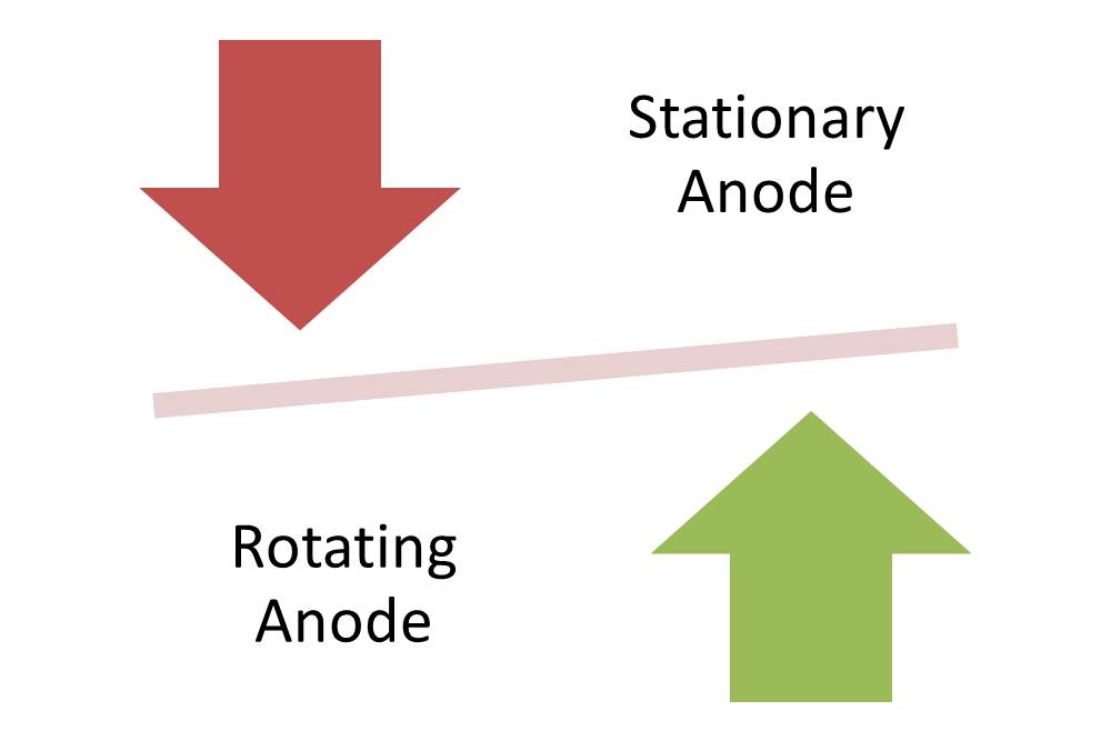
Most C-arms today come with dual focal spots. However, one may have to choose between stationary or rotating anode options. In a rotating anode tube, the heat generated by cathode beam is dispersed relatively across a larger area of the anode, as it rotates. This ensures longer life of the anode even if longer scans at higher doses are performed .
Rotating anode systems can be used for longer duration and at a higher dose. If in case large number of cases are likely to involve longer scans like run-offs or cross laterals, or scans requiring higher dose for larger patients, the anode is likely to get heated more. In this scenario, the superior heat dispersal of the rotating anode would be preferable option. Popular rotating anode models include the OEC 9600, 9800, Allengers HF 49R, or 59R.
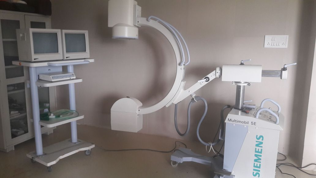
On the other hand, if mainly short, lower-dose studies, such as pain management needle placement or hand and foot specialties, a stationary anode tube would suffice. Models like the Siemens Compact L, Allengers HF 49, Philips Surgico 60-D HF, Siemens Multimobil 5C, RMS Corus C-arm are popular models with stationary anode.
Compact Monoblock C-Arm Systems
Most C-Arms today use Monoblock technology so as to reduce cost and size as compared to traditional tube plus generator configurations. This also eliminates the high voltage cabling connecting the tube and generator. As a result, Monoblock is more compact and cost-effective than traditional designs.
Collimator System
Iris Collimation, Virtual collimation or smart collimation with ability to move each leaf individually are all technology deployments mainly aimed at lower radiation exposure in the recent C-Arm models. Radiation exposure to the patient can be reduced through use of adjustable (iris) collimator to limit the field to the area of interest. This also improves image resolution.
FOV – Do I need a 6” Image Intensifier C-arm or 9” one?
An image intensifier (II) is an integral part of C-Arm machines, converting x-rays into high-intensity light forming brighter images as compared to just a fluorescent screen.
Image intensifiers come in a range of input field of view (FOV) diameters for diagnostic imaging applications, from 6 inches (15 cm FOV) to 16 inches (40 cm FOV), and many dimensions in between, depending on the type of imaging procedure, the C-arm is required for. While the higher II size/diameter provides with larger field of view, the lower II mode gives greater degree of magnification and clarity.
C-arms come with single-field, dual-field or triple-field image intensifiers (II). So, in a triple-field II, the 9″ image intensifiers could come in 4.5″, 6″ and 9″ size while the 12″ image intensifier could be in the 6″, 9″ and 12″ size. Older models like Siemens Multimobile 5C came with 6” and 9” dual modes.
For orthopedics, pain management or general surgery 9″ image intensifier would be suitable. Vascular procedures would generally need the 12″ image intensifier. The larger field of view of the 12″ magnification mode enables visualization of larger portion of the body enabling the surgeon to do procedures in a one-shot instead of seeking multiple views as may be the case if a 9” II was used.
C-Arm machine with 12″ image intensifiers also tend to be slightly larger in size as a unit, hence they need more space and are also costlier.
What image processing, display, storage and transfer facilities do I need?
Most C-Arm machines today come with high resolution CCD camera, two 17” or bigger monitors for display, storage of atleast 100 frames or more, as well as USB data port for transfer of images. Older systems like Siemens Multimobile 5C had only standard four frame image memory in addition to Last Image Held. It is important features to consider when buying a C-Arm machine to check the image handling capability of the C-arm depending on the way it will be used. If the C-Arm is likely to be used only during the procedure, with no need for future referral to the images or transfer and storage in a Hospital Information System or Electronics medical records of the patient, then one need not worry about the storage and transfer capacity.
Recent advances in C-Arm systems
Some of other key considerations for recent advances in C-Arm systems have focused around–
- Minimising Radiation dosage and exposure control
- Image quality, processing capabilities and quality retention over a period of time
- Maneuverability or Ease of movement e.g. isocentricity
Other features in advanced systems
Radiation dosage and exposure reduction
Airkerma display – The total Airkerma for each Patient at the normal Patient distance from X-ray source is displayed along with Dose per procedure, Dose rate and Dose Area Product on the Live Monitor. While purchasing a C-Arm with budgetary constraints, it is a common tendency to go for the cheapest possible machine which can deliver results for the basic applications required. Minimizing radiation leakage and dosage requirements seem to be nice-to-have stuff. But the physicians and surgeons must remember that it is not only the adverse impact on the patient, but even themselves and the supporting staff who are constantly exposed to higher levels of radiations than necessary. This is an important features to consider when buying a C-Arm machine.
- Flouroscopy modes – Modern fluoroscopy systems based on image intensifiers are extremely flexible devices. These can be operated in a wide range of modes for dynamic and static imaging. Accompanying this flexibility is the fact that different imaging modes have different dose characteristics. Fluoroscopes typically have the capability of operation in a number of dynamic imaging modes: normal fluoroscopy, high-dose fluoroscopy, and conventional and digital cine fluoroscopy. In addition, these systems may record analog or digital static images (eg, conventional photospot images, digital photospot images). Modes such as pulse fluoroscopy modes enable good quality images with minimum effective exposure to radiation.
- Laser aimer/ marker – A laser aimer is an additional optional tool provided in some of the C-arms. Laser aimer is used in spot identification, better alignment or c-arm positioning, without multiple exposures.
3D imaging
C-arm computed tomography is a new and innovative imaging technique. It uses two-dimensional (2D) X-ray projections acquired with a FDP C-arm system to generate images like CT slices. These images are visualized either as cross-sectional images or as 3D data sets using different volume rendering in combination with 2D fluoroscopic or radiographic imaging. 3D C-arm imaging provides valuable information for therapy planning, guidance, and outcome assessment all in the interventional suite.
Flat-panel detectors (FDP)
FDPs are increasingly replacing image intensifiers (II) on mobile C-arm systems. This is an important features to consider when buying a C-Arm machine. The advantages of a flat panel detector include: lower patient dose and increased image quality. There is no deterioration of the image quality over time. With FDP, the overall physical size of the C-arm is also reduced significantly as compared to usage of image intensifier.
Display and image resolution
Display and image resolution is getting more and more sophisticated in advanced systems. While most basic C-Arms have image acquisition through CCD camera and 512 matrix display, advanced systems have HD (1K x 1K), FHD and UHD high resolution displays. Most C-arms have 2 X 17” monitors for live and static images. Some of the high end systems have 32” UHD TV systems. Image storage options range from 10,000 to 300000 images. USB ports for image transfer is common, advanced system feature DICOM compatibility and DVI-I outputs.
User Interface
Touch screen operated systems, multiple operational control such as foot-switch as well as hand-held control, anti-glare monitors, easy to sterilize modules, On-screen virtual keyboard and image control keypad and several other convenient user interface features are part of advanced systems. Pre-set imaging profiles are also very convenient for users.
Image manipulation
Advanced systems provide for advanced image processing capabilities. Images processing such as zoom, pan, flip, invert, torch and median, Image rotation on both active and passive monitors when offline are all available in some systems. Snapshot feature produces Digital Radiography image quality in fluoroscopy mode. Image Annotations feature is helpful to record and communicate the observations.
PrimedeQ is an eMarketplace for medical equipment. We offer all types of imaging equipment including C-Arm machines. We also assist in medical equipment repair & maintenance services at www.Primedeq.com, including assistance for AERB licensing. Contact us at +918971223957 or +917019759765 for all your medical equipment related needs.
https://in.linkedin.com/in/shanthi-mathur-ab07838